|
|
|
|
|
Navigation links
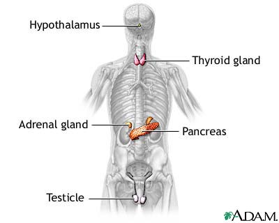
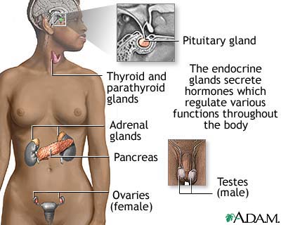
The endocrine system helps regulate and maintain various body functions by synthesizing (making) and releasing hormones, chemical messengers. The major areas of control and integration include responses to stress and injury, growth and development, absorption of nutrients, energy metabolism, water and electrolyte balance, reproduction, birth, and lactation. The endocrine system is composed of glands that release their hormones directly into the bloodstream for chemical signaling of target cells. These glands include the pituitary gland, the pineal gland, the hypothalamus, the thyroid gland, the parathyroid glands, the thymus, the adrenal glands, the ovaries (in females) or testes (in males), and the pancreas.
Typically, the body synthesizes hormones in one part and transports it to another through the bloodstream or lymph. Endocrine glands have a rich blood supply through which hormones travel to reach their target organs. Hormones alter the metabolism of target organs by increasing or decreasing their activity. These changes in activity are strictly balanced to maintain homeostasis (a stable internal environment). Glands are of two types. Endocrine glands do not have a duct system and are called ductless glands. These glands release hormones directly into the blood or lymph. Exocrine glands such as the sudoriferous (sweat) glands contain ducts. Ducts are tubes leading from a gland to its target organ.
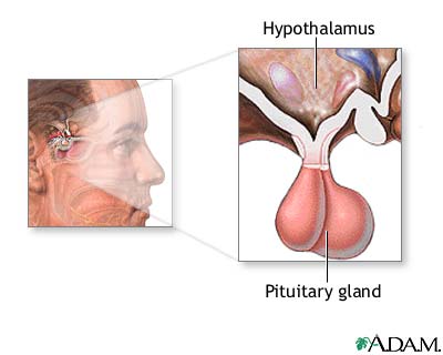
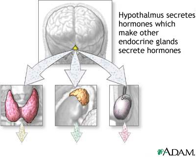
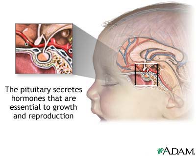
The endocrine system and the nervous system are so closely associated that they are collectively called the neuroendocrine system. Neural control centers in the brain control endocrine glands. The main neural control center is the hypothalamus, also known as the "master switchboard." Suspended from the hypothalamus by a thin stalk is the pituitary gland. The hypothalamus sends messages to the pituitary gland; the pituitary gland, in turn, releases hormones that regulate body functions.
Homeostatic feedback mechanisms Many endocrine glands are linked to neural control centers by homeostatic feedback mechanisms. The two types of feedback mechanisms are negative feedback and positive feedback. Negative feedback decreases the deviation from an ideal normal value, and is important in maintaining homeostasis. Most endocrine glands are under the control of negative feedback mechanisms. Negative feedback mechanisms act like a thermostat in the home. As the temperature rises (deviation from the ideal normal value), the thermostat detects the change and triggers the air-conditioning to turn on and cool the house. Once the temperature reaches its thermostat setting (ideal normal value), the air conditioning turns off.
An example of negative feedback is the regulation of the blood calcium level. The parathyroid glands secrete parathyroid hormone, which regulates the blood calcium amount. If calcium decreases, the parathyroid glands sense the decrease and secrete more parathyroid hormone. The parathyroid hormone stimulates calcium release from the bones and increases the calcium uptake into the bloodstream from the collecting tubules in the kidneys. Conversely, if blood calcium increases too much, the parathyroid glands reduce parathyroid hormone production. Both responses are examples of negative feedback because in both cases the effects are negative (opposite) to the stimulus. Positive feedback mechanisms control self-perpetuating events that can be out of control and do not require continuous adjustment. In positive feedback mechanisms, the original stimulus is promoted rather than negated. Positive feedback increases the deviation from an ideal normal value. Unlike negative feedback that maintains hormone levels within narrow ranges, positive feedback is rarely used to maintain homeostatic functions. An example of positive feedback can be found in childbirth. The hormone oxytocin stimulates and enhances labor contractions. As the baby moves toward the vagina (birth canal), pressure receptors within the cervix (muscular outlet of uterus) send messages to the brain to produce oxytocin. Oxytocin travels to the uterus through the bloodstream, stimulating the muscles in the uterine wall to contract stronger (increase of ideal normal value). The contractions intensify and increase until the baby is outside the birth canal. When the stimulus to the pressure receptors ends, oxytocin production stops and labor contractions cease. The pea-size pituitary gland is called the "master gland" because it regulates many key functions. The pituitary gland has an adenohypophysis (anterior lobe) and a neurohypophysis (posterior lobe). The adenohypophysis produces and secretes seven hormones in response to commands from the hypothalamus:
The TSH, ACTH, FSH, and LH hormones are tropic hormones that simulate other endocrine glands. In response, the other endocrine glands produce hormones that affect metabolism. For example, TSH from the pituitary gland stimulates the thyroid gland to produce thyroid hormones. In turn, thyroid hormones inhibit the release of calcium in the blood. Other adenohypophysis hormones have unique effects upon metabolism. ACTH acts upon the cortex (outer area) of the adrenal gland to produce steroid hormones. FSH and LH act upon women and men in regulating various sexual characteristics. Prolactin and growth hormone act upon certain body tissues; they do not affect specific organs. Prolactin travels to the breast tissue glands of nursing mothers, causing milk production. Growth hormone stimulates protein synthesis and cell division in cartilage and bone tissue. Gigantism results when excessive amounts of growth hormone are produced during childhood. Pituitary dwarfism occurs when too little growth hormone is produced. Acromegaly occurs when too much GH is produced during adulthood.
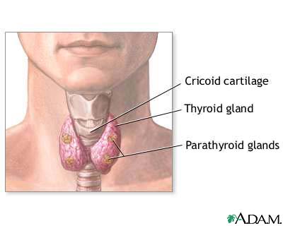
Neuron cell bodies of the hypothalamus produce two hormones: antidiuretic hormone (ADH) and oxytocin. These hormones are transported along the axons to the axon terminals in the pituitary posterior lobe. Both hormones are stored in the terminals until they are released into the blood vascular network surrounding the posterior pituitary gland. ADH acts upon the kidney tubules to help maintain a constant level of body water. This level is accomplished by increasing the water reabsorption amount when body water levels are low. Oxytocin triggers milk release from breast tissue when infants nurse and causes muscle contractions in the uterus during labor. The thyroid gland has two lobes connected by an isthmus (small connecting stalk) and is in the lower part of the neck just below the larynx. The thyroid gland produces three hormones:
T3 and T4 are collectively called thyroid hormone and are produced in the follicles (hollow spherical structures) of the thyroid gland. Thyroid hormone affects body growth, metabolic rates, and the development of bones and skeletal muscle. Thyroid hormone also increases the sensitivity of the cardiovascular system to sympathetic nervous activity. This effect helps maintain a normal heart rate. Parafollicular cells (C cells) between the thyroid gland follicles produce calcitonin. Calcitonin lowers blood calcium levels. The parathyroid glands are embedded in back of the thyroid gland and secrete PTH (parathyroid hormone). PTH increases blood calcium by stimulating bone calcium release into the bloodstream and by increasing the calcium absorption rate in the gastrointestinal tract and kidneys.
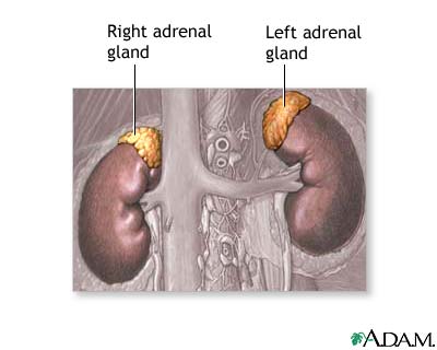
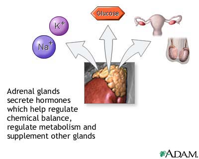
The adrenal glands are on top of each kidney. Each gland has a cortex (outer region) and a medulla (inner region). The cortex secretes glucocorticoids such as cortisol, mineralocorticoids, and small amounts of androgens and estrogens responsible for some secondary sex characteristics. Glucocorticoids raise blood sugar levels by increasing gluconeogenesis (synthesis of glucose from amino acid). This action ensures glucose supplies for the body when it is under stress. Mineralocorticoids such as aldosterone promote sodium (salt) reabsorption by stimulating the kidneys to absorb more sodium from the blood.
The medulla "emergency gland" develops from nervous tissue; the autonomic nervous system controls its secretions. The medulla secretes epinephrine (adrenaline) and norepinephrine (noradrenaline), chemicals that raise the blood levels of sugar and fatty acids. These hormones also increase the heart rate and force of contraction. These effects prepare the body for the "Fight or Flight" response (instant physical activity), enabling the individual to think quicker, fight harder, and run faster. These hormones also constrict the blood vessels supplying the skin, kidneys, gastrointestinal tract, and other areas of the body not needed for the response.
The ovary is the site of estrogen and progesterone synthesis. Estrogen is required to form the ovum (egg) during oogenesis and prepares the uterus for implanting a fertilized egg. Progesterone prepares the breasts for lactation during pregnancy and works with estrogen to regulate the menstrual cycle. The testes produce the hormone testosterone. Testosterone is required for sperm formation during spermatogenesis, the development of male external genitalia, and secondary sexual traits such as beard growth, chest hair, and enlarged thyroid cartilage. |
Any duplication or distribution of the information contained herein is strictly prohibited. The information provided herein should not be used for diagnosis or treatment of any medical condition and is provided for your general information only.