|
|
|
|
|
Navigation links All the body cells metabolically consume oxygen, and discharge carbon dioxide. To cover this need, respiration takes place internally (at the cellular level) and externally (ventilation/breathing). Ventilation involves the inhalation of atmospheric air into the lungs via the nose and mouth through branching passageways, and the exhalation of carbon dioxide. The lung key function is to bring air and blood into intimate contact in the alveolar air sacs so that oxygen can enter the blood, and carbon dioxide can leave. At rest, humans breathe about twelve times a minute, bringing in approximately a pint of air. Exercise and certain diseases result in a marked increase of breathing. The respiratory system also is vital in maintaining normal blood pH and body temperature.
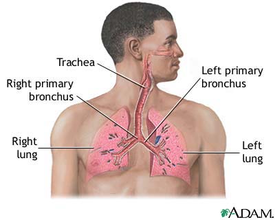
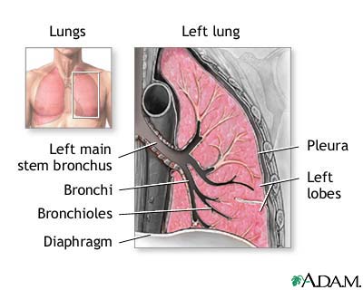
The lungs are paired organs that lie on either side of the heart and fill up the thoracic (chest) cavity. Inferior to (below) the lungs is the diaphragm, a broad thin muscle that separates the thoracic cavity from the abdominal (gut) cavity. On the medial (inner) surface of each lung is the hilus, where blood vessels, nerves, and bronchi (air passages) enter the lungs. The lungs differ in size and shape. Because the heart is slightly larger on the left side, the left lung has a cardiac notch (indented border). The left lung is also slightly smaller than the right. Each lung is divided into lobes (partitions) by fissures. The right lung has three lobes: lower, middle, and upper. These horizontal and oblique fissures create these lobes. The left lung has upper and lower lobes that are divided by the oblique fissure. Air enters the body through the mouth or nose. In the nose, thick hairs lining the nostrils prevent small objects from entering the nasal cavity. This cavity is lined with cells that produce mucus. Small foreign matter that enters the nasal cavities is trapped in the mucus, while tiny cilia (small hair-like projections) push the mucus to the pharynx (throat), where it is swallowed and digested in the stomach or expectorated.
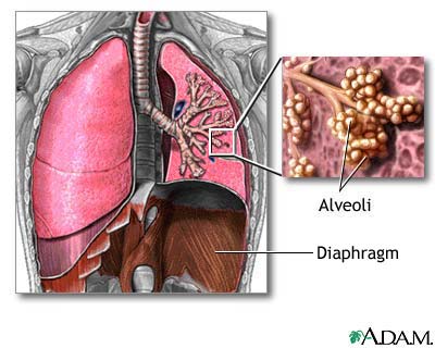
From the pharynx, the air passes to the larynx, which is called the voice box because it contains the vocal cords. To prevent food or liquid from entering the larynx, the epiglottis (a small flap of tissue) closes over the opening of the larynx during deglutition (swallowing). If this process works improperly, a cough reflex expels the foreign material. When air travels past the larynx, it enters the trachea (windpipe). The trachea is a strong tube containing rings of cartilage that prevents it from collapsing. The mucosa that lines the airway warms and moistens the air before it reaches the trachea. Within the lungs, the trachea branches into a left and right bronchus, which divide into increasingly smaller branches called bronchioles. The smallest bronchioles end in a cluster of air sacs, collectively called an acinus. The acinus comprises individual air sacs called alveoli. Alveoli are like small balloons that inflate and deflate with air during respiration.
Gas exchange occurs in the lungs between the alveoli and a capillary network within the alveolar wall. Capillaries are microscopic blood vessels that exchange material between the blood and body tissues. In the lung capillaries, blood from tissues where cellular metabolism is occurring is called deoxygenated blood because it contains many carbon dioxide molecules and few oxygen molecules.
The respiration process has two parts: inspiration (inhaling) and expiration (exhaling). During inspiration, the diaphragm contracts, moves downward, and causes the thoracic cavity volume to increase. Because the lungs are closely associated with the interior chest wall, they expand as the thoracic cavity expands. When the diaphragm relaxes (upward position), the thoracic volume decreases and the lungs partially deflate. This process is called expiration. The elastic recoil of the expanded thoracic wall and lungs also helps expiration.
After inhalation, the alveoli contain many oxygen molecules. The alveoli are in close contact with the capillary network. This proximity enables the minuscule oxygen molecules to diffuse (pass freely) from the alveolus to the bloodstream, flowing from a region of higher concentration to a region of lower concentration. In the bloodstream, the oxygen attaches to red blood cells and is transported to the rest of the body. Likewise, carbon dioxide diffuses from the bloodstream into the alveolus where it is transported out of the body during exhalation.
During respiration, the pleurae (pleural membranes) help the lungs to expand and contract. These membranes are sacs that tightly cover the lungs and the chest inside wall. Between these two linings is a space called the pleural cavity that contains a thin layer of fluid. This fluid allows the lungs to move freely against the thoracic cavity inside. |
Any duplication or distribution of the information contained herein is strictly prohibited. The information provided herein should not be used for diagnosis or treatment of any medical condition and is provided for your general information only.


