|
|
|
|
|
Navigation links
This system has 206 bones and associated cartilage, tendons, and ligaments. Because bone is rigid, it gives the body a framework, maintains its shape, and protects vital organs. Bones provide a place for muscles and supporting structures to attach, and, with the movable joints, form a system of levers upon which muscles can act to produce body movements. A joint is a place of union between two or more bones that may be movable or immovable. Bone also functions as a site for mineral storage and blood cell formation. Tendons and ligaments are strong bands of fibrous connective tissue that attach muscles to bones, and bones to bones, respectively.
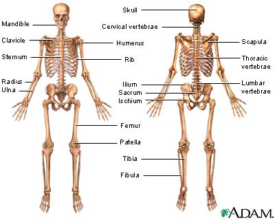
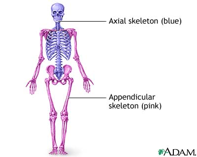
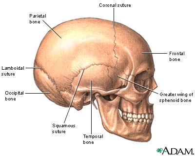
The skeleton has two parts: the axial skeleton and the appendicular skeleton. The axial skeleton includes the skull, the hyoid bone, the vertebral column (spine, sacrum, and coccyx), the sternum, and the ribs. Its components are aligned along the long axis of the body. The appendicular skeleton includes the bones of the upper extremities (arms, forearms, and hands), the pectoral (shoulder) girdle, the pelvic (hip) girdle, and the bones of the lower extremities (thigh, knee, leg, and foot). Its components are outside the body main axis. Bone tissue stores and releases ionic minerals that affect homeostasis (stable internal environment) of the body. Chief among these minerals is calcium, which is necessary for proper functioning of the muscles and nervous system. Endocrine system hormones regulate calcium release and storage. Cranial bones are flat, rounded, and fused to protect the brain. The eight cranial bones are the frontal (1), parietal (2), temporal (2), occipital (1), sphenoid (1), and ethmoid (1). Two cranial bones meet at a suture (immovable joint). The four major sutures are the coronal suture (between the parietal and frontal bones), the lambdoidal suture (between the occipital and parietal bones), the sagittal suture (between the two parietal bones), and the squamous suture (between the parietal and temporal bones).
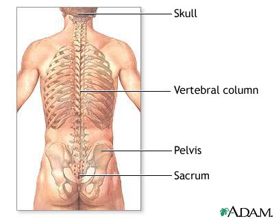
Facial bones provide a framework for the facial muscles, form eye sockets, and form jaws for the teeth. The fourteen facial bones are the vomer (1), maxilla (2), zygomatic (2), palatine (2), lacrimal (2), nasal (2), inferior nasal conchae (2), and mandible (1). The hinged mandible (jaw bone) moves freely during mastication (chewing) and speech. The vertebral column has three groups of vertebrae and two sets of fused bones. These vertebrae include seven cervical (neck) vertebrae, twelve thoracic (upper back) vertebrae, and five lumbar (lower back) vertebrae. Five fused vertebrae form the sacrum and from three to five fused small vertebrae form the coccyx (tail bone). The vertebrae form a column of bone that protects the spinal cord. The thoracic vertebrae have facets (indentations) upon their surfaces that articulate (meet) with the ribs. The twelve pairs of ribs are long, flattened, and curved bones that form a protective cage for the heart, lungs, and other internal organs. The vertebrosternal (true) ribs are the first seven ribs; they are "true" because they attach directly to the sternum (breast bone). Ribs eight through twelve are the false ribs because they indirectly attach to the sternum or they lack a sternal attachment. Ribs eight through ten are the vertebrochondral ribs because they attach indirectly to the sternum by cartilage. Ribs eleven and twelve are called floating (vertebral) ribs because they do not attach to the sternum. Instead, their floating position allows them to bend sideways while providing protection for the kidneys. In the appendicular skeleton, the pectoral girdle bones include two scapulae (shoulder blades) and two clavicles (collar bones). The scapula is in the upper back and articulates with two bones: the humerus and the clavicle. Because the scapula is part of the shoulder joint, the scapula must be mobile to allow the upper extremities freedom of movement. The clavicle articulates with the sternum and the scapula, giving support to the pectoral girdle and adding stability to the shoulder joint. The bones of the upper extremities are the humerus, ulna, radius, carpals (wrist bones), metacarpals (hand bones), and phalanges (finger bones). The humerus in the arm articulates with the scapula at the shoulder joint and with the ulna and radius at the elbow. The radius and ulna in the forearm articulate with the carpals at the wrist. The ulna articulates with the humerus and forms the elbow. The carpals are small, flat, irregularly shaped wrist bones. They articulate with the metacarpals in the hand. The metacarpals articulate with the phalanges. In the pelvic girdle are two hip bones. Each coxa (hip bone) forms from fused bones. The two coxae, sacrum, and coccyx form the pelvis, a bowl-shaped cavity that supports and protects many abdominal organs. The three fused bones of each coxa are the ilium, ischium, and pubis.
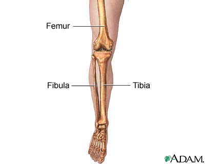
The pelvic girdle articulates with the femur (thigh bone) at the acetabulum (hip joint) and with the sacrum at the sacroiliac joint. Also, the coxae articulate with each other at the pubic symphysis, a joint with limited movement. The femur is the longest and heaviest bone in the body and helps in weight-bearing while standing. At the knee, the femur articulates with the tibia (shin bone). Suspended within muscle tendons at the front of the knee joint is the patella (kneecap). The patella is an example of a sesamoid bone (small bone) that is within a tendon. Attaching to the lateral (outer side) of the tibia is the fibula (leg bone). The fibula provides points of attachment for muscles of the foot and leg and increases the lateral stability of the ankle. The fibula is not a weight-bearing bone like the tibia. In the foot are specialized bones designed for weight-bearing. Among these are the tarsals (ankle bones), metatarsals (foot bones), and phalanges (toe bones). These foot bones form a system of arches that allow the foot to support much weight.
Ossification and reconstruction Ossification (bone growth) begins during fetal development. Before birth every skeleton bone appears as a fibrous membrane template (membranous bone) or a cartilaginous template (cartilaginous bone). These templates form the basic shapes that mature bone replaces. Membranous bone develops through intramembranous ossification. Cartilaginous bones develop through endochondral ossification. Despite its hardness and relative inflexibility, bone is one of the most changing tissues of the body. At the bone tissue cellular level, reorganization and replacement of the solid matrix occurs continuously. Two types of cells are primarily responsible for this reconstructive process: the osteoblasts and the osteoclasts.
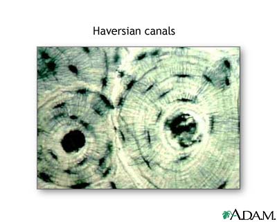
Osteoblasts form new bone tissue in response to more demands on bone. When a person is physically active, the rate of osteoblast activity increases, which thickens and strengthens the bones. When muscles tense and flex, their tendons pull on the bones. This action causes the osteoblasts to secrete proteins that form lamellar (compact) bone and cancellous (spongy) bone with texture that resembles trabeculae (a web-like network of tunnels). Within compact bone is a system of canals that contain blood vessels, nerves, and lymph vessels. These canals form functional units called osteons (Haversian systems). The osteon is a tube-like structure containing a central (Haversian) canal through which nerves and blood vessels pass. Surrounding the central canal is bone matrix, a substance produced by osteocytes. Within the osteon, osteocytes form lamellae, concentric layers of hard bone matrix. As osteoblasts create lamellae, these osteoblasts become trapped within the compact bone and are called osteocytes.
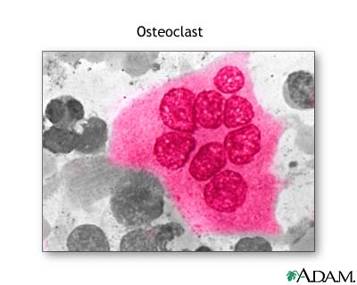
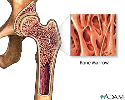
Osteocytes lie within lacunae, small pockets between the lamellae. Canaliculi, tiny canals, connect the lacunae. Canaliculi also connect with the central canal. These connections allow nutrients to pass through the canaliculi to the trapped osteocytes within the dense lamellae. Osteoclasts work with osteoblasts. These osteoclasts dissolve bone tissue by secreting enzymes. Their actions help healthy individuals keep bones light and airy. They also regulate calcium and phosphate concentrations in body fluids. When osteoclast activity is not balanced by new bone formation, osteoporosis, a degenerative bone disease, occurs. Within the long bones are two types of bone marrow: red marrow and yellow marrow. The yellow marrow has fatty connective tissue and fills the marrow cavity. During starvation, the body uses the fat in yellow marrow for energy.
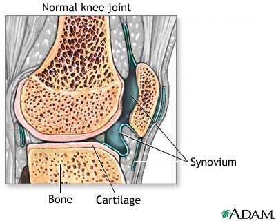
The red marrow of some bones is an important site for blood cell production. Here all erythrocytes (red blood cells), platelets, and most leukocytes (white blood cells) form in adults. From the red marrow, erythrocytes, platelets, and leukocytes migrate to the blood to do their special tasks. Red blood cells carry oxygen and nutrients to the body tissues. Platelets help in blood clotting. White blood cells help fight disease and infection. Bones of the skeleton articulate at joints. Joints form three categories of movability: freely movable, slightly movable, and immovable. Freely movable joints are also called synovial joints. A typical synovial joint has a joint capsule, a synovial membrane, synovial fluid, a joint cavity, and articular cartilage. A joint capsule surrounds the joint, supporting and stabilizing it. The synovial membrane is within the joint capsule. This membrane closely surrounds the joint and forms a joint cavity. The synovial membrane secretes synovial fluid that lubricates the articular surfaces of the joint. In some joints, the synovial membrane extends outside the joint capsule to form a bursa. The bursa cushions the joint. Bursae are in the knee, elbow, shoulder, and hip. Articular cartilage covers the articular surfaces of synovial joints to prevent excess wear and tear as they move against each other.
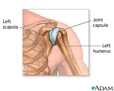
Six types of synovial joints are hinge, ball-and-socket, pivot, condyloid (angular or ellipsoidal), plane (gliding), and saddle. The elbow is an example of a hinge joint. Here, the convex and concave articulating bones allow movement along one plane, similar to a door. The shoulder and hip are the only ball-and-socket joints in the body. In this type of joint, one bone has a spherical head that articulates with a corresponding concavity. This joint frees the joint to move in many directions. In a pivot joint, one round-shaped articulating bone fits within a corresponding depression on another bone. This joint allows one bone to rotate against the other. An example is the radioulnar joint (joint of the radius and ulna) in the forearm. In a condyloid (angular) joint, one bone has an oval articulating head that rests within an oval concavity. This joint permits angular movement of the bones. The metacarpophalangeal joint (junction between the metacarpals and phalanges) of the hand are examples of condyloid joints.
Plane joints have two flat bones joined. The sole movement of the bones is short gliding motions. An example of this joint is the intertarsal joint (junction between the tarsal bones) of the feet. Saddle joint bones have convex and concave surfaces similar to a saddle. This joint allows the joint to move in many directions. The carpometacarpal joint of the thumb is an example saddle joint. As their name implies, amphiarthrosis joints (slightly movable joints) have limited movement. The two types of amphiarthrosis joints are syndesmosis (fibrous) and symphysis (cartilaginous). A syndesmosis joint occurs when two bones join by a section of cartilage. The junction between the tibia and fibula is an example. A symphysis joint forms when two bones fuse by a fibrocartilage pad. Typical symphysis joints are between the pubic symphysis (pubic bones in the pelvis), and in the vertebral column between individual vertebrae. Intervertebral discs act as weight-bearing shock absorbers for walking, jumping, and lifting. An immovable joint is called a synarthrosis. The two types of this joint are sutures and gomphoses. Sutures are joined by short fibers of dense fibrous connective tissue and are in the skull. The single example of a gomphosis joint is the teeth sitting within their sockets. An example of a bony fusion joint is the fusion of the three bones forming a coxa (hip bone): the ilium, ischium, and pubis. Ligaments link bones. These ligaments, sheets of strong, fibrous connective tissue, are identical with the muscular system tendons. The only difference is their function: tendons attach muscle to bone and ligaments attach bone to bone. |
Any duplication or distribution of the information contained herein is strictly prohibited. The information provided herein should not be used for diagnosis or treatment of any medical condition and is provided for your general information only.